Departamento de Fisiologia e Farmacologia, Departamento de Clínica Odontológica, Departamento de Química Orgânica e Inorgânica, Universidade Federal do Ceará, Fortaleza, CE, Brasil
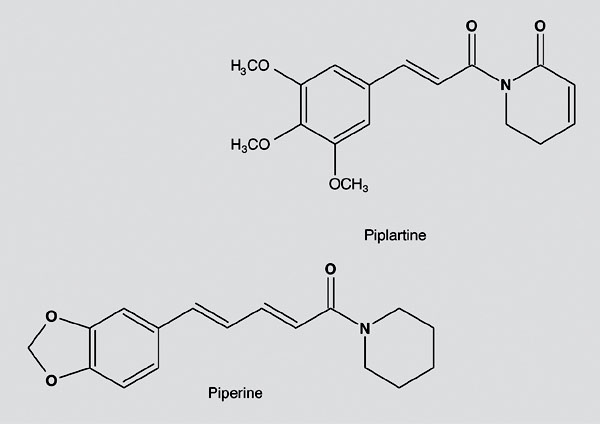
Piplartine {5,6-dihydro-1-[1-oxo-3-(3,4,5-trimethoxyphenyl)-2-propenyl]-2(1H)pyridinone} and piperine {1-5-(1,3)-benzodioxol-5-yl)-1-oxo-2,4-pentadienyl]piperidine} are alkaloid amides isolated from Piper.
Both have been reported to show cytotoxic activity towards several tumor cell lines. In the present study, the in vivo antitumor activity of these compounds was evaluated in 60 female Swiss mice (N = 10 per group) transplanted with Sarcoma 180. Histopathological and morphological analyses of the tumor and the organs, including liver, spleen, and kidney, were performed in order to evaluate the toxicological aspects of the treatment with these amides. Administration of piplartine or piperine (50 or 100 mg kg-1 day-1 intraperitoneally for 7 days starting 1 day after inoculation) inhibited solid tumor development in mice transplanted with Sarcoma 180 cells. The inhibition rates were 28.7 and 52.3% for piplartine and 55.1 and 56.8% for piperine, after 7 days of treatment, at the lower and higher doses, respectively. The antitumor activity of piplartine was related to inhibition of the tumor proliferation rate, as observed by reduction of Ki67 staining, a nuclear antigen associated with G1, S, G2, and M cell cycle phases, in tumors from treated animals. However, piperine did not inhibit cell proliferation as observed in Ki67 immunohistochemical analysis. Histopathological analysis of liver and kidney showed that both organs were reversibly affected by piplartine and piperine treatment, but in a different way. Piperine was more toxic to the liver, leading to ballooning degeneration of hepatocytes, accompanied by microvesicular steatosis in some areas, than piplartine which, in turn, was more toxic to the kidney, leading to discrete hydropic changes of the proximal tubular and glomerular epithelium and tubular hemorrhage in treated animals.
Introduction
Modern man is confronted with an increasing incidence of cancer and cancer deaths annually. Statistics indicate that men are largely plagued by lung, colon, rectal, and prostrate cancer, while women increasingly suffer from breast, colon, rectal, and stomach cancer[1]. The literature indicates that many natural products are available as chemoprotective agents against commonly occurring cancer types[2].
In addition to their use as spices, Piper species (Piperaceae) are important plants in traditional medicine. In particular, they are useful against asthma, bronchitis, fever, hemorrhoidal afflictions, gastrointestinal diseases, and rheumatism[3]. The preparations obtained from plants belonging to the genus Piper have shown anti-inflammatory, antifeedant, insecticidal, and anti-hypertensive activities, as well as inhibitory activity of aflatoxin B DNA binding and anticancer activity[3]. Many investigators have emphasized that the phytochemicals isolated from spices such as pepper species are potent biological agents with interesting anticancer properties[4].
Piplartine and piperine are alkaloid-amide components obtained from Piper species. Several of them have been reported to show cytotoxic activity towards tumor cell lines[5]. Piperine has been reported to display central nervous system depressant, antipyretic, analgesic, and anti-inflammatory activities[3]. Piperine also protects against chemical carcinogen-induced genotoxicity[6]. Moreover, piperine is a potent inhibitor of the mixed function oxygenase system and of p450 isoenzymes[7]. Piperine has been reported to inhibit the in vitro and in vivo production of nitric oxide and tumor necrosis factor-a[8], and to inhibit lung metastasis[9]. Pradeep and Kuttan[10] demonstrated that piperine is also a potent inhibitor of nuclear factor-kß (NF-kß) and proinflammatory cytokine gene expression in B16F-10 melanoma cells, which, according to these investigators, could explain its anti-metastatic activity. Sunila and Kuttan[11] also demonstrated piperine’s immunomodulatory and antitumor activities.
Piplartine, on the other hand, has been much less studied. Its in vitro antiproliferative activity was demonstrated using tumor cell lines[12][13][14]. Recently, Bezerra et al.[14] compared the antimitotic activity of piperine and piplartine in different cell systems, e.g., in cultured tumor cells and sea urchin embryos, and concluded that piplartine was more potent.
In the present study, we evaluated the antitumor activity of piplartine and piperine in mice transplanted with Sarcoma 180. Histopathological and morphological analyses of the tumor and the organs, including liver, spleen and kidney were performed in order to evaluate the toxicological aspects of the treatment with these amides.
Material and Methods
Plant material
The roots of Piper tuberculatum were harvested in September 2004 from a wild population on the Pici Campus of the Federal University of Ceará, Fortaleza, CE, Brazil. A voucher specimen (#34736) was deposited in the Prisco Bezerra Herbarium (Escola de Agronomia do Ceará), Department of Biology, Federal University of Ceará.
Piplartine isolation
Ground roots of P. tuberculatum (450 g) were macerated with a 1:1 mixture of petroleum ether:ethyl acetate (1.5 L) for 24 h (three times). The solvent mixture was evaporated under reduced pressure to yield a yellowish solid (13.24 g), which, after crystallization from MeOH, provided a first crop of piplartine (4.35 g; Figure 1). The melting point was 122.2-122.6ºC (Lit. 128-130º and 124ºC) and piplartine was also characterized by 1-D and 2-D NMR[15][16].
Piperine from black pepper seeds was purchased from Acros Organics (Fair Lawn, NJ, USA)
Figure 1 – Chemical structure of piplartine and piperine

Figure 1. Chemical structure of piplartine {5,6-dihydro-1-[1-oxo-3-(3,4,5-trimethoxyphenyl)-2-propenyl]-2(1H)pyridinone} and piperine {1-5-(1,3)-benzodioxol-5-yl)-1-oxo-2,4-pentadienyl]piperidine}.
Assay of antitumor activity
Sixty female Swiss mice weighing 20-30 g, obtained from the central animal house of the Federal University of Ceará, Brazil, were used. Animals were housed in cages with free access to food and water. All animals were maintained under a 12-h light-dark cycle (lights on at 6:00 am).
Sarcoma 180 tumor cells were maintained in the peritoneal cavities of mice from the same animal house. Ten-day-old Sarcoma 180 ascites tumor cells (2 x 10⁶ cells/500 µL) were implanted subcutaneously into the left hind groin of the experimental mice. One day after inoculation, piperine and piplartine (50 and 100 mg/kg) or 5-fluorouracil (5-FU, 25 mg/kg) were dissolved in 10% DMSO and administered intraperitoneally for 7 days. The negative control was injected with 10% DMSO. On day 8, the mice were sacrificed and tumors, livers, spleens, and kidneys were excised, weighed and fixed in 10% formaldehyde. The inhibition ratio (%) was calculated by the following formula: inhibition ratio (%) = [(A – B)/A] x 100, where A is the average tumor weight of the negative control, and B is the tumor weight of the treated group.
Histopathology and morphological observations
After fixation in formaldehyde, tumors, livers, spleens, and kidneys were grossly examined for size or color changes and hemorrhage. Then, portions of the tumor, liver, spleen, and kidney were cut into small pieces, followed by staining with hematoxylin and eosin of the histological sections. Histological analyses were performed by light microscopy, with priority given to recognizing the extent of liver or kidney lesions attributable to the drugs.
Immunohistochemical Ki67 detection
Tumor sections were deparaffinized with xylene and dehydrated with ethanol. The slides were then immersed in water for 10 min. For antigen retrieval, the slides were boiled in citrate buffer, pH 6.0, for 15 min in a microwave oven and subsequently cooled for 20 min. Next, the slides were washed in TBS and endogenous peroxidase was blocked with 0.3% hydrogen peroxide for 15 min. After washing with TBS, the sections were incubated overnight at 4ºC with mouse antibodies to Ki67 at 1:50 concentration. After 24 h, the slides were washed and incubated with a multilink antibody for 20 min, washed in TBS and incubated for 20 min with the avidin-biotin-peroxidase complex. After washing with TBS, the slides were incubated for 3 min with diaminobenzidine, and finally counter-stained with hematoxylin prior to mounting. The percentage of proliferating neoplastic cells was evaluated directly by light microscopy. To quantify the amount of proliferation, all Ki67-positive cells were counted in 4-6 random fields per slide.
Statistical analysis
Data are reported as mean ± SEM. The differences between experimental groups were compared by ANOVA followed by the Student-Newman-Keuls test, with the level of significance set at P < 0.05.
Results
The effects of piplartine and piperine on Sarcoma 180 tumors transplanted in mice are shown in Table 1. A significant reduction of tumor weight was observed at both doses both in piplartine- and piperine-treated animals (P < 0.05). On day 8, the average tumor weight was 1.96 ± 0.13 g in control mice and 1.41 ± 0.11 and 0.94 ± 0.16 g in mice treated with piplartine at the dose of 50 and 100 mg kg-1 day-1, respectively. In the presence of piperine, weights were 0.89 ± 0.20 and 0.85 ± 0.12 g at the same doses. The tumor growth inhibition ratios were 28.7-52.2 and 55.0-56.8% for piplartine and piperine, respectively. The inhibitions were significant for both compounds and doses in relation to the control group (P < 0.05). Nevertheless, the effect of piplartine was clearly dose-dependent (P < 0.05), while for piperine there was no significant differences between the two doses used (P > 0.05). With the dose of 25 mg kg-1 day-1 5-FU reduced tumor weight by 76.7%, within the same period.
After treatment with piperine (100 mg kg-1 day-1) and 5-FU (25 mg kg-1 day-1), the spleen weights were significantly reduced (P < 0.05), while piplartine treatment did not induce any change in the organs’ weight (Table 1). Histopathological analysis of the kidneys from treated animals showed discrete hydropic changes of the proximal tubular epithelium and glomerular and tubular hemorrhage, but the glomerular structure was essentially preserved. The alterations seemed to be more intense in animals treated with piplartine. In addition to the effects observed in the kidneys, the histopathological analyses indicated that the liver was also a target organ for piperine and piplartine toxicity, with the effects of piperine being more intense than those of piplartine. These effects included Kupffer cell hyperplasia, portal tracts and centrolobular venous congestion, focal infiltrate of chronic inflammatory cells, intense ballooning degeneration of hepatocytes, microvesicular steatosis, and sinusoidal hemorrhage. However, in the presence of piplartine, no areas of steatosis were observed.
Histopathological analysis of the tumors excised from control mice showed groups of large, round and polygonal cells, with pleomorphic shapes, hyperchromatic nuclei and binucleation. Several degrees of cellular and nuclear pleomorphism were seen. Mitosis, muscle invasion and coagulation necrosis were also noticed. In the tumors excised from animals treated with 5-FU, piperine and piplartine, extensive areas of coagulative necrosis were observed.
Ki67 staining for cell proliferation was performed in the tumors removed on day 8 from untreated animals and from animals treated with 5-FU (25 mg kg-1 day-1), piperine and piplartine (100 mg kg-1 day-1). Figure 2 shows the amount of Ki67-positive cells on the slides analyzed. The relative number of Ki67-positive tumor cells was significantly reduced in tumors from mice treated with 5-FU (94.3% inhibition) and piplartine (45.1% inhibition) compared to control tumors (P < 0.05) or tumors treated with piperine.
Figure 2 – Effect of antitumor substances on the cell proliferation of Sarcoma 180 cells

Figure 2. Effect of antitumor substances on the cell proliferation of Sarcoma 180 cells. The mice received 25 mg kg-1 day-1 5-fluorouracil (5-FU) and 100 mg kg-1 day-1 piperine and piplartine for 7 days. Ki67-positive cells from 4-6 fields/tumor were counted. Data are reported as the mean ± SEM number of positive cells/field for 10 mice in each group. *P < 0.05 compared to control (ANOVA followed by the Student-Newman-Keuls test).
Table 1 – Effect of piplartine and piperine on Sarcoma 180 tumors transplanted in mice
Discussion
The present paper reports the antitumor activity of piplartine and piperine on mice transplanted with Sarcoma 180. Piplartine and piperine are alkaloid-amide components from Piper species. Piplartine and piperine have been reported to show antiproliferative effects in vitro, with piplartine being more potent than piperine[12][13][14]. Sarcoma 180 is a mouse-originated tumor and one of the most frequently used cell lines in antitumor-related research in vivo[17][18][19]. Both piplartine and piperine presented activity in this model. The in vivo antitumor activity of piperine has been previously demonstrated in Dalton’s lymphoma ascites model[11]; however, the present report is the first one on the in vivo antitumor activity of piplartine.
According to Sunila and Kuttan[11], the antitumor activity of piperine is related to its immunomodulatory properties, which involve the activation of cellular and humoral immune responses. Piperine also had an inhibitory effect on lung metastases[9], probably due to NF-kß inhibition and proinflammatory cytokine gene expression[10]. In fact, piperine possessed only weak cytotoxic activity[10][11][14], indicating that its antitumor activity is not related to direct antiproliferative effects on tumor cells.
Immunohistochemical staining of cells for proliferation-associated proteins provides information about the tumor proliferation rate. The monoclonal antibody Ki67, described by Gerdes et al.[20], is a mouse monoclonal antibody that identifies a nuclear antigen associated with the G1, S, G2, and M phases. This molecule is expressed along the entire cell cycle, except in G0 and early G1[21]. Thus, the results obtained with Ki67 staining support the notion that piperine activity was not related to a reduction of tumor proliferation rate.
The mechanisms involved in the antitumor activity of piperine have not yet been elucidated. Moreover, its immunostimulant activity still remains controversial, since Dogra et al.[22] demonstrated that piperine at the dose of 4.5 mg/kg had a consistent immunosuppressive effect, suppressing the weight and cell population of the spleen of treated animals. In the present study, we also observed a reduction of spleen weight in piperine-treated animals, but only in animals treated with the 100 mg/kg dose.
The histopathological analyses suggested that the liver is the main affected organ in piperine treatment. Ballooning degeneration of hepatocytes, accompanied by microvesicular steatosis in some areas, was observed in the group treated with piperine, suggesting intrinsic hepatotoxicity, which produces liver damage when the drug is taken in sufficient quantities. However, regeneration of hepatic tissue occurs in many diseases, except in the most deleterious ones. Even when hepatocellular necrosis is present, but connective tissue is preserved, regeneration is almost complete[23][24]. The hepatic alterations observed after piperine treatment could be considered reversible[23][24][25].
Piplartine, on the other hand, is considered to be a strong cytotoxic alkaloid, inhibiting the proliferation of several tumor cell lines in culture[12][13][14]. These data suggest that the presence of two a,ß-unsaturated carbonyl moieties is an important structural requirement for cytotoxic activity[12][13][14].
In fact, the activity profile of piplartine seems to be different from that of piperine. Immunohistochemical results obtained with Ki67 staining showed that the antitumor activity of piplartine is associated with a reduction in the tumor proliferation rate, which is compatible with its antiproliferative effects. Piplartine had no effect on the spleen of treated animals, and apparently has the kidney as the main toxicological target. Moreover, the kidney damage observed in piplartine-treated animals could also be considered reversible[26].
The present data show that both piplartine and piperine possess in vivo antitumor activity related to reversible toxic effects on liver and kidney. Thus, piplartine and piperine may act as antitumor agents, although they seem to act through different pathways and the mechanisms of action responsible for their tumor-reducing activity are not known.
Acknowledgments
The authors thank Silvana França dos Santos and Maria de Fátima Teixeira for technical assistance.
Correspondence and Footnotes
Address for correspondence: L.V. Costa-Lotufo, Departamento de Fisiologia e Farmacologia, UFC, Rua Cel. Nunes de Melo, 1127, 60430-270 Fortaleza, CE, Brasil. Fax: +55-85-4009-8333. E-mail: lvcosta@secrel.com.br.
Research supported by CNPq, Instituto Claude Bernard, FUNCAP, Banco do Nordeste, and FINEP. Received August 25, 2005. Accepted March 24, 2006.
References & External links
- Abdulla M, Gruber P. Role of diet modification in cancer prevention. Biofactors 2000; 12: 45-51.
- Reddy L, Odhav B, Bhoola KD. Natural products for cancer prevention: a global perspective. Pharmacol Ther 2003; 99: 1-13.
- Parmar VS, Jain SC, Bisht KS, Jain R, Taneja P, Jha A, et al. Phytochemistry of the genus Piper. Phytochemistry 1997; 46: 597-673.
- Lampe JW. Spicing up a vegetarian diet: chemopreventive effects of phytochemicals. Am J Clin Nutr 2003; 78: 579S-583S.
- Tsai IL, Lee FP, Wu CC, Duh CY, Ishikawa T, Chen JJ, et al. New cytotoxic cyclobutanoid amides, a new furanoid lignan and anti-platelet aggregation constituents from Piper arborescens. Planta Med 2005; 71: 535-542.
- Selvendiran K, Padmavathi R, Magesh V, Sakthisekaran D. Preliminary study on inhibition of genotoxicity by piperine in mice. Fitoterapia 2005; 76: 296-300.
- Atal CK, Dubey RK, Singh J. Biochemical basis of enhanced drug bioavailability by piperine: evidence that piperine is a potent inhibitor of drug metabolism. J Pharmacol Exp Ther 1985; 232: 258-262.
- Pradeep CR, Kuttan G. Effect of piperine on the production of nitric oxide and TNF-a in vitro as well as in vivo. Immunopharmacol Immunotoxicol 1999; 25: 337-346.
- Pradeep CR, Kuttan G. Effect of piperine on the inhibition of lung metastasis induced B16F-10 melanoma cells in mice. Clin Exp Metastasis 2002; 19: 703-708.
- Pradeep CR, Kuttan G. Piperine is a potent inhibitor of nuclear factor-kappaB (NF-kappaB), c-Fos, CREB, ATF-2 and proinflammatory cytokine gene expression in B16F-10 melanoma cells. Int Immunopharmacol 2004; 4: 1795-1803.
- Sunila ES, Kuttan G. Immunomodulatory and antitumor activity of Piper longum Linn. and piperine. J Ethnopharmacol 2004; 90: 339-346.
- Duh CY, Wu YC, Wang SK. Cytotoxic pyridone alkaloids from Piper aborescens. Phytochemistry 1990; 29: 2689-2691.
- Duh CY, Wu YC, Wang SK. Cytotoxic pyridone alkaloids from the leaves of Piper aborescens. J Nat Prod 1990; 53: 1575-1577.
- Bezerra DP, Pessoa C, de Moraes MO, Silveira ER, Lima MA, Elmiro FJ, et al. Antiproliferative effects of two amides, piperine and piplartine, from Piper species. Z Naturforsch [C] 2005; 60: 539-543.
- Braz-Filho R, Souza MP, Mattos MEO. Piplartine-dimer A, a new alkaloid from Piper tuberculatum. Phytochemistry 1981; 20: 345-346.
- Atal CK, Dhar KL, Singh J. The chemistry of Indian Piper species. Lloydia 1975; 38: 256-264.
- Ito H, Shimura K, Itoh H, Kawade M. Antitumor effects of a new polysaccharide-protein complex (ATOM) prepared from Agaricus blazei (Iwade strain 101) “Himematsutake” and its mechanisms in tumor-bearing mice. Anticancer Res 1997; 17: 277-284.
- Lee YL, Kim HJ, Lee MS, Kim JM, Han JS, Hong EK, et al. Oral administration of Agaricus blazei (H1 strain) inhibited tumor growth in a sarcoma 180 inoculation model. Exp Anim 2003; 52: 371-375.
- Kintzios SE. What do we know about cancer and its therapy. In: Kintzios SE, Barberaki MG (Editors), Plants that fight cancer. Boca Raton: CRC Press; 2004. p 1-14.
- Gerdes J, Schwab U, Lemke H, Stein H. Production of a mouse monoclonal antibody reactive with a human nuclear antigen associated with cell proliferation. Int J Cancer 1983; 31: 13-20.
- Gerdes J, Lemke H, Baisch H, Wacker HH, Schwab U, Stein H. Cell cycle analysis of a cell proliferation-associated human nuclear antigen defined by the monoclonal antibody Ki-67. J Immunol 1984; 133: 1710-1715.
- Dogra RK, Khanna S, Shanker R. Immunotoxicological effects of piperine in mice. Toxicology 2004; 196: 229-236.
- Scheuer PJ, Lefkowitch JH. Drugs and toxins. In: Scheuer PJ, Lefkowitch JH (Editors), Liver biopsy interpretation. London: WB Saunders; 2000. p 134-150.
- Kummar V, Abbas A, Fausto N. Pathologic basis of disease. Philadelphia: WB Saunders; 2004.
- McGee JOD, Isaacson PA, Wright NA. Oxford textbook of pathology: pathology of systems. New York: Oxford University Press; 1992.
- Tisher CC, Brenner BM. Renal pathology: with clinical and functional correlations. Philadelphia: JB Lippincott Company; 1994.
The original text taken from a:
![]() http://www.scielo.br/scielo.php?pid=S0100-879X2006000600014&script=sci_arttext
http://www.scielo.br/scielo.php?pid=S0100-879X2006000600014&script=sci_arttext
Bezerra, D. P., et al. "In vivo growth-inhibition of Sarcoma 180 by piplartine and piperine, two alkaloid amides from Piper." Brazilian journal of medical and biological research 39.6 (2006): 801-807.
Available under conditions of the license:![]() http://creativecommons.org/licenses/by/4.0/deed.en
http://creativecommons.org/licenses/by/4.0/deed.en


Comments
“In vivo growth-inhibition of Sarcoma 180 by piplartine and piperine, two alkaloid amides from Piper”